
When conducted in person, the Ramzi Theory boasts an impressive accuracy rate of 97%. This method relies on analyzing the placement and orientation of the chorionic villi (future placenta) to determine the sex of your baby.

Baby Gender Prediction: Ramzi Theory explained by Ultrasound Technicians
This article covers Dr. Ramzi’s ultrasonography Research to explain how to use this method when you are approximately 6-8 week’s pregnant and go to your first ultrasound. If you are further along in your pregnancy (closer to 12-14 weeks), try using the Nub Theory and/or Skull Theory for early gender prediction.
“Ramzi’s method uses the placenta/chorionic villi location as a marker for fetal gender detection at 6 weeks gestation, and it was found to be highly reliable. The Ramzi method correctly predicts the fetus’ gender in 97.2% of the males and 97.5% of the females early in the first trimester.” obgyn.net
Dr. Saam Ramzi Ismail conducted an extensive and controlled study of over 5,000 pregnant women. He discovered that using the direction or orientation of the chorionic villi (future placenta) is an accurate marker in determining the sex of a baby. Scientifically, it relates to a natural polarization in which male embryos have a proven pattern of being magnetized toward the right side of the uterus, and females are drawn toward the left side.
Chorionic villus form before 9 weeks’ gestation. It turns into the placenta which provides oxygen and nutrients to the fetus and removes waste products from the baby’s blood. The Mayo Clinic describes the chorionic villi as “wispy projections of placental tissue that share the baby’s genetic makeup.”
Boy or Girl? How to read the ultrasound scan when using the Ramzi Method
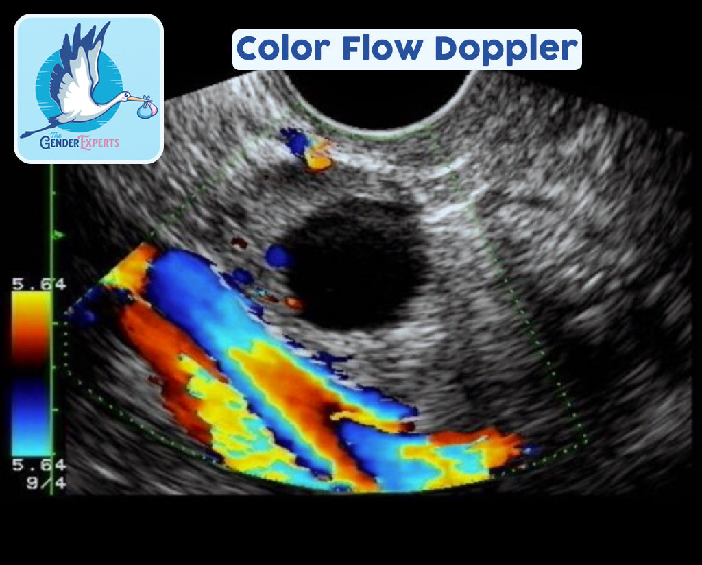
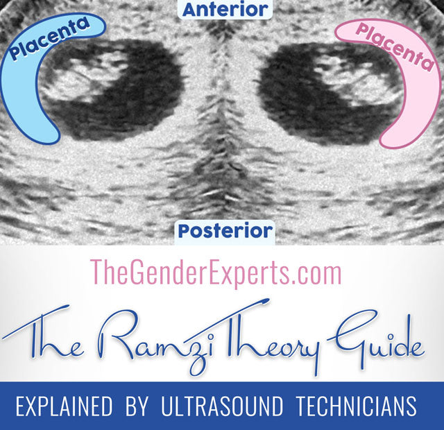
Dr. Ramzi's study adhered to rigorous guidelines and incorporated a control group to obtain reliable results. Utilizing color flow Doppler technology to identify the direction and pinpoint the location of the chorionic villi yields a high level of accuracy in determining the baby's sex. Requesting your technician to employ this technology increases the likelihood of obtaining precise results. When the chorionic villi or future placenta are situated on the left side of your body, it is indicative of a probable girl. Conversely, if they are located on the right side of your body, it is more likely to be a boy!
The identification of the chorionic villi, which will eventually become the placenta, is done by locating a bright white area on the ultrasound scan. Detecting the placenta may be easier in some scans compared to others. In certain cases, the scan may reveal multiple bright white areas. In medical terminology, "hyperechoic" refers to a characteristic of material that produces echoes of higher amplitude or density than the surrounding medium. It can also describe a region in an ultrasound image where the echoes are stronger than normal or surrounding structures. When examining your ultrasound photo to locate the placenta or yolk sac, focus on the areas with higher density.
A transvaginal ultrasound, also known as an endovaginal scan, differs from an abdominal ultrasound as it involves inserting the ultrasound wand approximately 2 or 3 inches inside the vaginal canal to capture internal images. These images can provide a view of the vagina, cervix, uterus, fallopian tubes, and even the ovaries. Internal scans are commonly used in early pregnancy to obtain clear and high-quality visuals for the doctor or technician. In a transvaginal ultrasound, the technician can determine the location of the placenta on both the left and right sides of the uterus. This information is helpful when applying the Ramzi Method for gender prediction. However, there is a debate among pregnant women discussing the Ramzi Theory regarding whether transvaginal ultrasounds reflect the same orientation as depicted in the scan, as abdominal scans are typically "flipped" or mirror images. Due to this discrepancy, it is important to note that not all scans are equally suitable for the Ramzi Theory. While abdominal ultrasounds are generally mirrored, there are cases where they are not, making them consistent with the same maternal side.
Technicians can help determine the correct orientation
When applying the Ramzi Method, The Gender Experts, examine the following aspects of the scan:
- Annotations present on the scan, such as “TRV,” “TRAN,” “LONG,” “R/L Flip,” or “SAG.”
- Surrounding anatomy, including the presence of ovaries, cervix, bladder, and other structures.
- The shape of the sac, which may require rotation by 30, 90, 180, or other degrees clockwise or counterclockwise to aid in analysis.
Products
Select a Product and upload your scan. Our experts will review and deliver The Gender Prediction Report same day, directly to your E-mail.
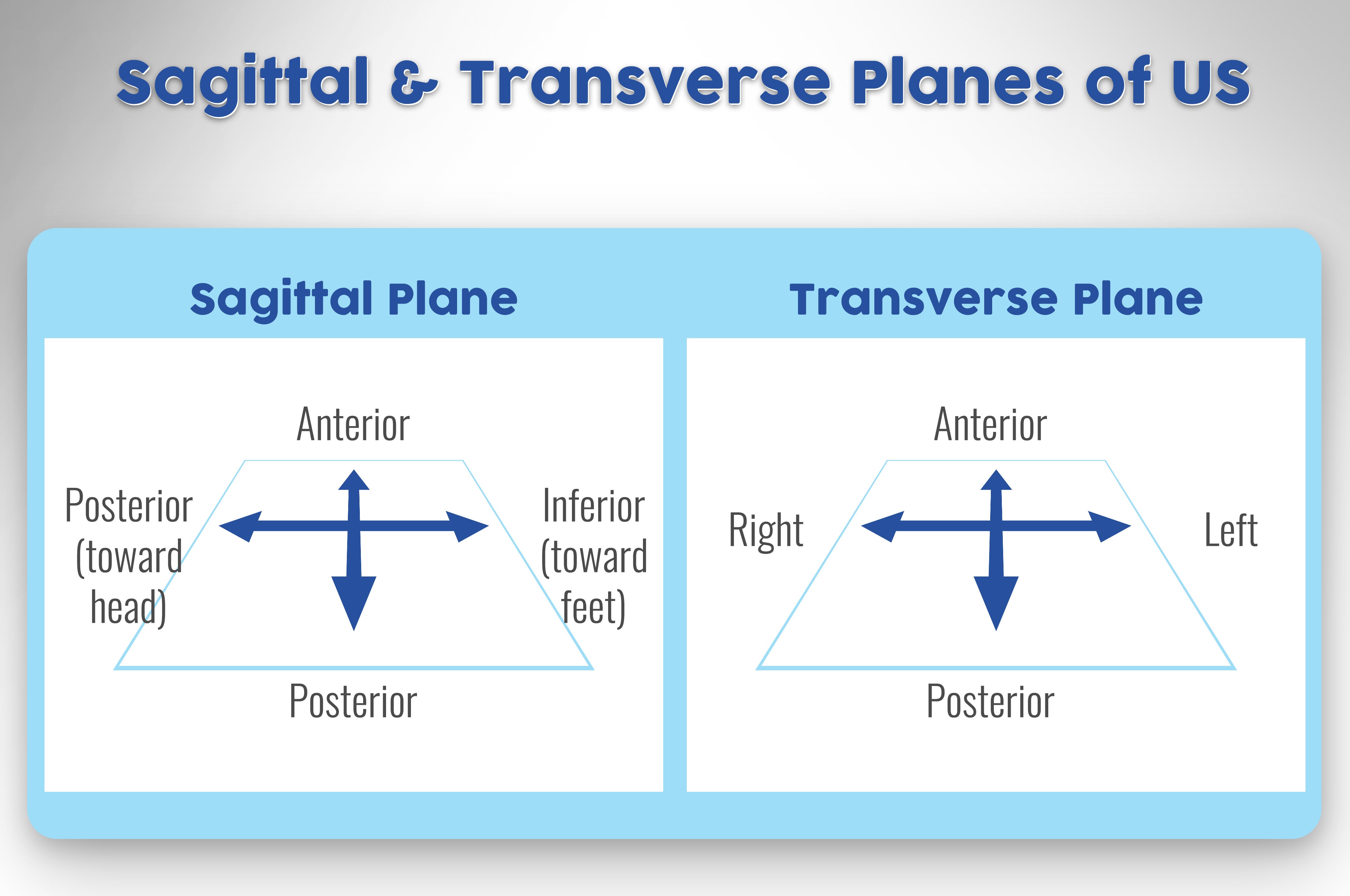
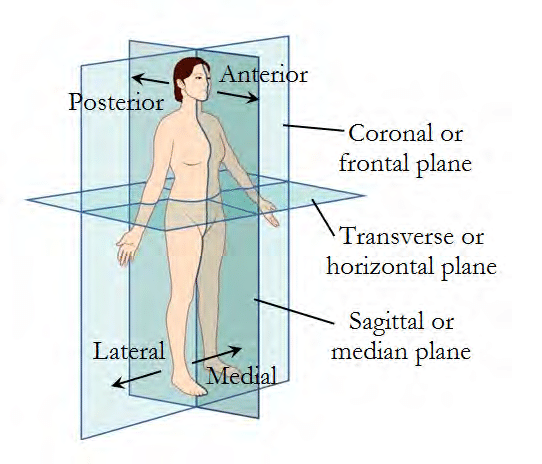
Sagittal Scanning Plane
A sagittal scanning plane provides a view that appears to slice from left to right, as if looking from the right side of your belly towards the center. While this view allows for identifying the orientation of up and down or front and back, it doesn't provide information about right versus left. It is commonly referred to as a "long view," and the only determination that can be made regarding placenta location in this view is whether it is anterior (toward the front) or posterior (toward the back). Due to this limitation, sagittal scanning planes can negatively impact the accuracy of the Ramzi Theory.
Transverse Scanning Plane
On the other hand, for accurate determination of the side where the future placenta is located, Ramzi Theory scans should be performed in the transverse plane. The transverse plane is akin to looking from your belly button inward, allowing for clear visualization of the left and right sides of the uterus. This specific scanning plane is preferred to ensure reliable results when applying the Ramzi Theory.


Ramzi Method Example Pictures:
Using ultrasound photos as a guide for predicting the gender of your baby can be a fun and entertaining activity. However, it's important to recognize that there is always a possibility of error in such predictions. If you are uncertain or seeking a higher accuracy rate, submitting your Ramzi Theory scan to us, or asking your technician for assistance can provide more reliable results. The correct positioning of the placenta is a crucial factor in determining whether you are carrying a boy or a girl. While the Ramzi Theory offers a higher level of accuracy compared to traditional old wives' tales, it's important to note that online gender predictor methods are primarily intended for entertainment purposes.
Confirmed Scans Gallery
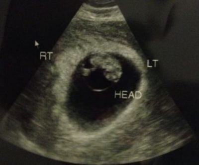
This ultrasound image confirms a male fetus at 8 weeks' gestation. Note that the baby is positioned in the center. However, according to the Ramzi Theory, the determination of gender is based on the location of the placenta, specifically the chorionic villi at this stage. In this picture, the future placenta is observed on the left side of the image or the right side of the body. To view more confirmed Ramzi scans, please visit our Confirmed Scans Gallery. Another clue for gender prediction is the position of the yolk sac, as it is typically close to the chorionic villi. In this scan example, the yolk sac leans toward the left side of the image (right side of the body).
Additional evidence supporting a male fetus is the decidual reaction observed in the scan, which is also located on the right side of the body. The decidual reaction refers to the thickening of the endometrium seen in early pregnancy. The presence of a double decidual sac sign is one of the initial indications of pregnancy.

Ramzi Theory Frequently Asked Questions
In cases where twins have their own individual placentas, which is the majority of the time, the Ramzi Theory can potentially be used to predict the sex of each twin. However, it is important to note that there haven't been enough studies conducted specifically on using the Ramzi Theory for twins.
If you have ultrasound photos of your twins, you can send them to TheGenderExperts.com via our Shop. Our team of experts will be delighted to examine the photos and analyze the presence of yolk sacs, chorionic villi, and the locations of the placentas in relation to the rest of your uterus. They will do their best to provide insights and determinations based on the available information.
No. The side from which a woman's ovary releases an egg does not necessarily correlate with the location where the baby implants in her body. It is possible for a woman to release an egg from her left ovary, but the baby may implant on the right side of her body, and vice versa. Additionally, it's important to note that many women have only one functioning ovary, which means they have an equal chance of conceiving a male or female baby, regardless of which ovary the egg is released from. Therefore, the side of ovulation does not have any correlation with the side on which the placenta will form.
The Ramzi Theory allows for the prediction of the fetus's sex as early as 5 or 6 weeks' gestation, even before the formation of the placenta. During your initial pregnancy ultrasound, when confirming your pregnancy, you can request a transverse plane scan and ask the technician to mark the right and left sides of your uterus on the screen or photo. Using this information, you can make a prediction about the sex of your fetus.
While it is possible to make predictions using a later ultrasound scan, it's important to note that the accuracy may be affected. As the placenta grows in size, it becomes more challenging to determine which side it initially formed on. Therefore, the early ultrasound scans provide a more reliable basis for applying the Ramzi Theory and predicting the fetus's sex.
If it’s difficult for you to determine, send us your scan and we can help you!
More Gender Prediction Methods
If you haven’t had your ultrasound yet, or did not receive a picture, you can try some popular at-home Gender Prediction tests like the Gender Charts and Calendars or the simple and fun Baking Soda and Baby Heart Rate Gender Prediction Test.