
If you are pregnant, your health care provider will recommend an ultrasound exam at around week 20. This article covers what can you expect at the full anatomical ultrasound scan appointment.


What does the ultrasound scan show?
The ultrasound technology has improved significantly since it was invented. If the baby cooperates, you will be able to see spines, fingers, toes and his/her face. You may also be able to see the fetal gender.
It is important that you make your intentions clear about knowing the gender so that the technician doesn’t spill the beans. If you don’t inform the technician, then you might find out accidentally; or worse, you might not find out at all.
Although you might not be able to know much of what you are seeing on the ultrasound scan, your technician will be able to interpret it. The transducer is moved over your belly and your baby will also be moving around too.

Send your scans to our experts in ultrasonography for a same-day gender determination report delivered to your e-mail.
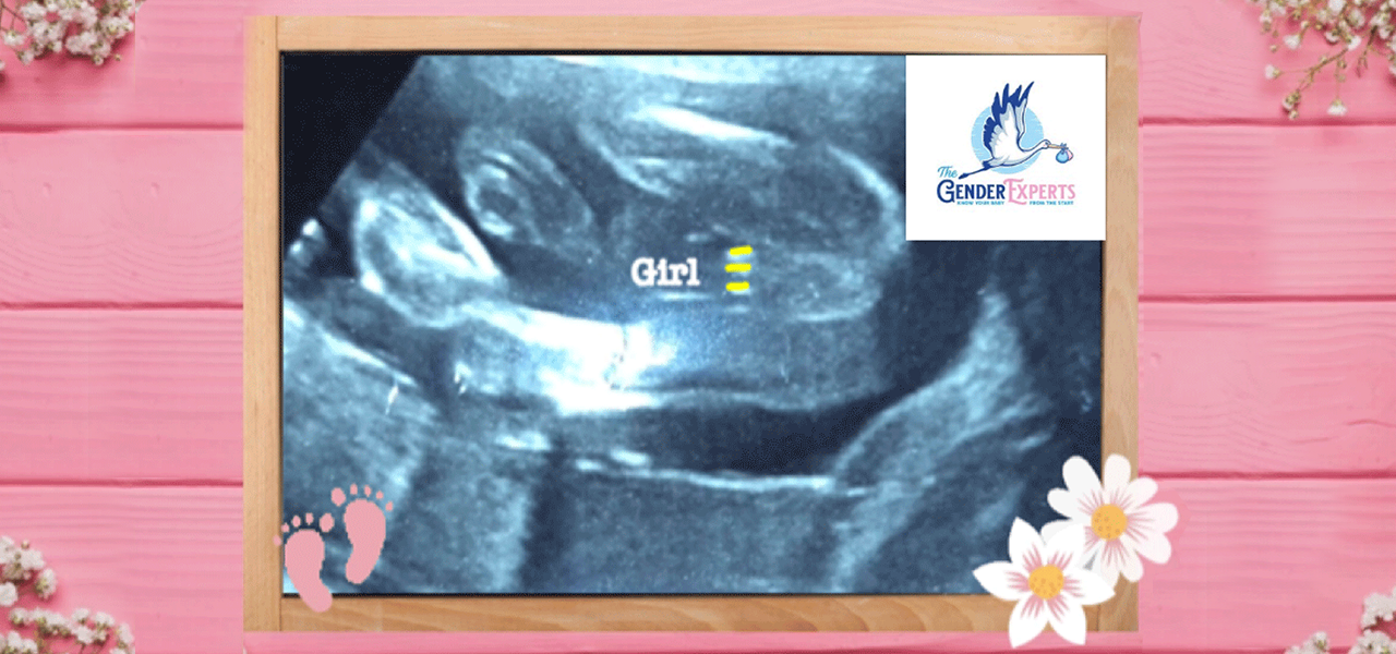
Girl Ultrasound Signs
The moniker given to girl parts appearance is the “hamburger sign.” The labia would be associated with the bun and the clitoris would resemble the hamburger patty. (3 white lines on the ultrasound) We have highlighted the 3 lines in yellow in our confirmed girl ultrasound scan pictured above. The sagittal sign is taken using a profile view. If there is a parallel downward pointing caudal notch (referred to as the nub theory in earlier stages of pregnancy), this is also female marker. From 12-14 weeks, it is roughly 90% accurate, and by 18 weeks, it is over 99% accurate.
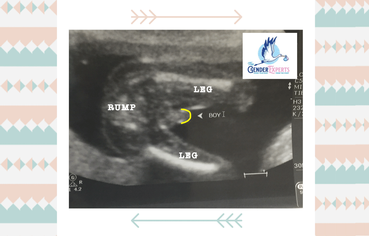
Boy Ultrasound Signs
Male genitalia is relatively easy to identify, especially during the exam when the technician observes the testicles, scrotum and penis. It is an obvious protrusion between the legs resembling a hot dog rather than a hamburger. The flow of urine is another “boy sign.” This can sometimes be spotted during your second trimester anatomy exam. If the urine flow is moving upward, then it is more than likely a male fetus. This is something only your technician would be able to show you. The Nub theory can also be used after 12 weeks gestation. The caudal notch would be upward facing and greater than 30’ in relation to the spine.
Other Fetal and Uterine Observations
Spine
Most women think their baby’s spine look like a string of pearls. It reminds you of the spines on dinosaur skeletons you see in a museum.
Face and skull
Waiting to see your baby’s face? At this stage, your baby’s face will look like a skull mask from different angles, but it will really feel nice to see your baby sucking his or her thumb. Learn more about the Skull Theory to assess the baby's gender for entertainment purposes.
Amniotic fluid:
Fluid will appear darker on the ultrasound scan and harder tissue like bone will appear bright.
Baby’s heart
Your technician will zoom in on your baby’s beating heart, looking at all four chambers, while also measuring the heart rate.
Crown to rump length
The sonographer will routinely measure from the top of the head to the rump to keep track of baby’s growth throughout your pregnancy. Your baby’s weight can also be estimated.
Placenta
Placenta is observed for its location (anterior or posterior), and whether there are any cysts, placenta previa or other abnormalities.
Fetal movement and position
Typical fetal movement at this stage show the baby in a relaxed position and regular movements. Since babies have a regular sleep and wake period, a lack of movement is not necessarily a cause for concern.
Other parts
The bladder, kidneys, umbilical cord and insertion point are routinely observed.
Preparing for Your Ultrasound Scan
There isn’t much to do when it comes to preparing for your ultrasound. The sonogram specialist will often recommend that you have a full bladder. You may want to carry a bottle of water so that you can drink as you sit in the waiting room.
Why do you need a full bladder? When the bladder is full, it pushes the uterus up and out of the pelvis a bit. This makes it easy to visualize the baby, amniotic sac, uterus and the umbilical cord.
Your anatomy scan will typically last 30-45 minutes. You may want to bring a loved one with you to share in the sneak peek of your little one. Some people like to record the session, especially if it will be the first time finding out the sex of the baby! Ask your provider prior to your session if it is permitted.